Right Atrium
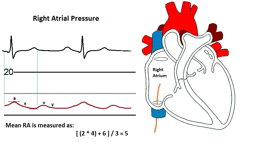
After the RV / LV matchup pressures are obtained and analyzed, the catheter is then pulled back into the Right Atrium (RA) across the Tricuspid valve. Notice how the waveform now mimics the PCWP or LA pressure waveform. It is generated from the same mechanical actions. Lets take a look at the waves of the right atrial waveform.
Components of the RA waveform
- The a wave is produced by right atrial systole and occurs after the P wave of the ECG.
- The a wave is followed by the x descent, reflecting right atrial relaxation.
- The c wave, not always seen, is produced by the closure of the tricuspid valve
- The v wave is produced by filling of the right atrium and occurs after the T wave of the ECG.
- The v wave is followed by the y descent, reflecting the opening of the tricuspid valve and the flow of blood into the right ventricle.
The RA is best viewed at a 40 or 50 mm Hg scale (dependent on what you monitor allows) at a speed of 25 mm/sec.
Notice the slight delay between electrical activity and pressure events. This is related to the distance between pressure transmitted from the right atrium through the catheter to the transducer. This length of your pressure tubing can contribute to this delay.
Always record the pressure at end expiration. Inspiration will create a decrease in intrathoracic pressure that will then lower the cardiac hemodynamic pressures.
The RA is recorded as: a/v/mean. Since the a wave and v wave are usually identical, the mean value is usually stated.
Mean pressure can be calculated as: [(2 * diastolic) + systolic] / 3.
Normal mean RA pressure is 2 - 6 mm Hg.
Reasons for elevated RA pressure
- RV Heart failure
- Tricuspid Valve Stenosis and/or Regurgitation
- Cardiac Tamponade
- Constrictive Pericarditis
- Pulmonary Hypertension
- Chronic Left Heart failure
- Volume overload
Just as mitral valve regurgitation produces elevated v waves in the LA / PCWP waveform, tricuspid valve regurgitation will produce elevated v waves in the RA waveform.
Reasons for low RA pressure
- Hypovolemia
- Shock
Right Atrial Blood Gas
One, or multiple, blood sample is pulled from the right atrium for analysis.
In normal situations, the RA oxygen saturation should be higher than the PA oxygen saturation. as blood flows toward the lungs, the oxygen saturation decreases before being oxygenated by the lungs. If a step-up, or increase in blood oxygenation is noticed, then a left-to-right shunt may need to be further evaluated. A left-to-right shunt is indicated with a step-up in the PA oxygen saturation is greater than 7% of the RA oxygen saturation. If a step-up is noticed, then multiple oxygen saturations from various points of the right atrium may be obtained.
It is important to remember that the coronary sinus empties into the right atrium. If a blood gas is obtained in the right atrium near the coronary sinus, the oxygen saturation will be abnormally high.
The normal right atrial blood gas oxygen saturation is 70 - 80%.
References
- Daily, E.K. and Schroeder, J.S. (1990) Hemodynamic Waveforms - Exercises in Identification and Analysis (2nd ed.). Philadelphia, PA: Mosby.
- Kern, M. et al (2003) The Cardiac Catheterization Handbook (4th ed.). Philadelphia, Pa: Mosby.