Vascular Access
Vascular access of the arterial system is essential for Left Heart Catheterzation and the assessment of the coronary arteries and left ventricle. Access of the venous system is essential for Right Heart Catheterization and the hemodynamic assessment of the heart and pulmonary vasculature
For the Left Heart Catheterizatin, femoral arterial access is the most widely used site. Other sites include the radial and brachial regions of the arm. Femoral arterial access is palpated just below the inguinal ligment.
Femoral Anatomy Review - Access should be obtained over the region of the femoral head. Important anatomical landmarks should be know regarding vascular access at the femoral site. The right groin anatomical sequence is: Nerve, Artery, anc Vein. The left groin anatomical sequence is: Vein, Artery, and Nerve.
After the arterial pulse is located, local anesthesia is given subcutaneously both superficially and into the deep tissue along both sides of the artery. Allow 2 to 3 minutes for local anesthesia to take effect.
Percutaneous puncture is then achieved with a hollow beveled tip needle advanced into the skin at a 30 - 45 degree angle. The needle is then advanced to the anterior wall of the artery. The pulse may be felt by the needle as it makes contact with the arterial wall. (A)As the needle enters the artery, pulsatile blood will be expelled from the proximal end of the needle. (B)The needle is then stabilized with one hand while the other hand advances the j-tipped guidewire through the needle and into the artery with a gentle action. If resistance is met with the wire, remove the wire and confirm pulsatile flow of blood from the needle.
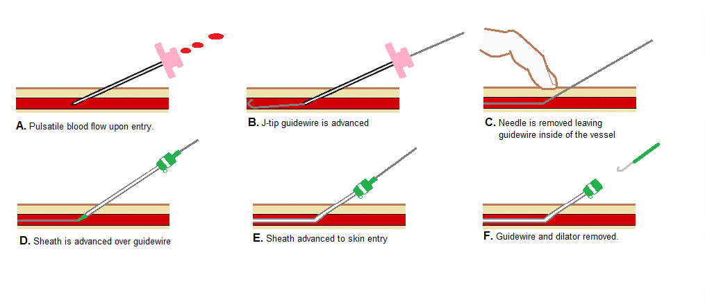
(C)With the j-tipped guidewire in the artery, the needle is removed, leaving the guidewire in the vessel. A small incision at the entry of the guidewire can be made with a scalpel to help with the insertion of the vascular sheath. (D)The sheath is then advanced over the guidewire and into the artery with clockwise and counter-clockwise rotations. (E)The sheath allows for a single arterial entry point for catheterization. (F)With the sheath in place, the guidewire and sheath dilator is removed, leaving the sheath inside the artery. Aspiration and flushing of the sheath with heparinized saline will prevent blood clot formation and embolization into the peripheral vasculature.
One of the complications associated with vascular access is a vasovagal reaction from the needle puncture and sheath manipulation. A vasovagal reaction usually results in bradycardia and hypotension. Treatment includes: IV Atropine, bolus of IV fluids, and oxygen supplementation.
References