Hemodynamics
Hemodynamics is the study of blood flow under external forces. There are several factors involved, but we want to focus on the cardiac chambers involved and what to look for. When reviewing hemodynamics, remember the principle of hydromechanics - fluid flows from regions of high pressure to regions of low pressure.
In the Cardiac Cath Lab, hemodynamics are seen in both Left Heart Catheterization (LV pressure and AO pressure) and Right Heart Catheterizatin (VC, RA, RV, PA, and PCWP). A Right Heart Catheterization is primarily performed to evaluate valvular heart disease, cardiac shunts and heart failure.
The upward and downward waves of hemodyanmic pressures are created by the increase and decrease of pressure in the vascular system. Waveforms are captured by fluid filled catheters that receive the pulsating pressure and then transmits that signal from the transducer to the monitor / recorder.
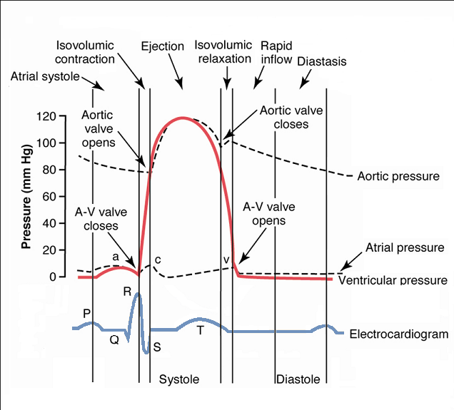
Before you begin your study of hemodynamics, understand the cardiac cycle and the pressures associated with each event. This will allow you to establish a foundation to further understand abnormalities in hemodynamics. The Wiggers diagram, by Dr. Carl J. Wiggers, is an excellent resource to understand the cardiac cycle. Take your time with this diagram. Look at each phase of the ECG and visualize what is happening with the heart and the change in waveform that is produced.
Set up Manifold for Recording Hemodynamics
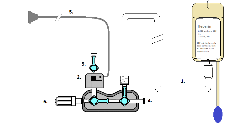
- Hand the saline spike tubing (1) off to circulator to attaching the manifold to a pressurized heparized saline bag. Flush the entire manifold to eliminate bubbles. Tap all of the connections to dislodge any air bubbles. Then close the stop cock off to pressurized saline bag.
- After the transducer (2) has been flushed, turn the stopcock (3) off to the transducer. The manifold stopcocks should be positioned to allow the transducer to be open to air (4) to establish a zero. When zeroing the transducer, it should be positioned at mid chest level of the patient.
- Hand transducer cable (5) off to circulator to connect to hemodynanmic monitoring system.
- Attach manifold (6) to fluid filled catheter to be used for hemodynamic recording.
- To obtain a waveform, turn stopcock arm to allow fluid signal to be transmitted to the transducer.
References
- Baim, D.S. (2005) Grossman's Cardiac Catheterization, Angiography, and Intervention. (7th ed.). Philadelphia,Pa: Lippincott, Williams & Wilkins.
- Kern, M. et al (2003) The Cardiac Catheterization Handbook (4th ed.). Philadelphia, Pa: Mosby.