Coronary Artery Disease
Coronary Artery Disease (CAD) is the narrowing of the coronary artery from atherosclerosis - the buildup of cholesterol and fatty deposits on the inner wall of the artery. This narrowing is often referred to as a blockage, lesion, or plaque and can restrict blood flow to the heart muscle.
CAD decelops when fat builds up along the endolthelial cellular wall of the artery. Other substances, such as inflammatory cells, cellular waste products, proteins and calcium can build up underneath the endothelial later of the arterial wall.
Over time, the inside of the arteries develop plaques of different sizes. As the plaque continues to develop, blood flow to the heart muscle becomes limited which becomes a problem during periods of increased demand. Diminished blood flow can lead to a period of ischemia that causes the individual to experience chest pain or angina. If the plaque surface cracks or ruptures, your body will will treat the vessel as it normally would to an injury to your body by sending platelets to that area. The platelets will cause blood cells to clot around the placque. This process can rapidly close the artery and completely block blood flow to the heart muscle.
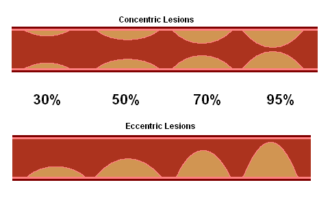
When we visualize the coronary angiogram, it is important to realize that we are only seeing the internal lumen of the artery in a 2-dimensional field. CAD is labeled as a lesion, stenosis, or blockage. The assessment of CAD includes type of lesion (concentric or eccentric) and the severity in relation to percentage of reduction in the internal lumen diameter.
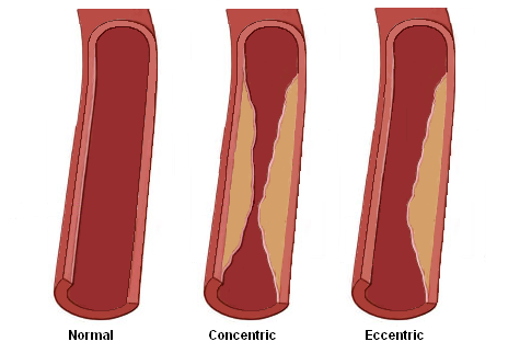
Concentric lesions are blockages with symmetric narrowing. These lesions have identical appearances in various projections.
Eccentric lesions are blockages with asymmetric narrowing and will have smooth and irregular appearances at different projections.
The diameter percentage of a lesion is estimated by viewing the vessel with the greatest narrowing. This percentage is scored by comparing it to the proximal and distal normal portions of the artery. Percentage is estimated by the naked eye and comes with experience. Imaging systems may contain programming that will analyze lesions and calculate percentages.