Left Coronary Angiography
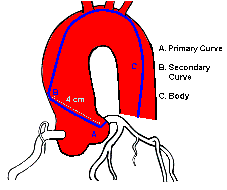
The most commonly used catheter for Left Coronary Angiography is the Judkins Left 4 cm curve (JL4) catheter. The 4 reflects the length of the segment between the primary and secondary curve of the catheter. See image below.
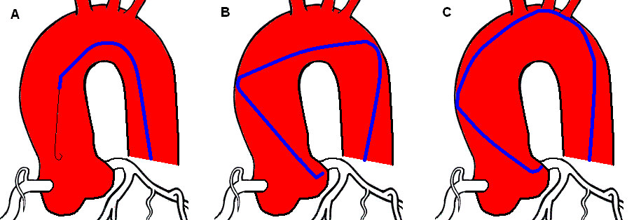
The JL4 is designed to engage the left coronary with very little manipulation. The best view to visualize the catheter cannulating the LCA is in the left anterior oblique (LAO) 30 to 50 degrees. With the tip of the catheter positioned just below the coronary ostium, the operator will give the catheter a gentle pull back allowing the catheter to fall into the LCA. If the Aorta is dilated, a longer JL catheter will need to be used (JL4.5 -6). Patients with a small or narrow aortic arch will usually need a JL3.5 catheter to cannulate the LCA. A catheter that is too short will have the tip pointed upward while a catheter that is too long will have the tip pointed downward.
Once the catheter is in the ostium of the LCA, IMPORTANT! observe the monitor for pressure dampening. If no dampening pressure is observed, then coronary injections can be administered. Multple views are taken of the left coronary to visualize the different branches. Refer to the angles below:
- LAO Caudal ("spider" view) - This angle provides excellent visualization of the Left Main Coronary Artdfy (LMCA) and how it bifurcates into the LAD and Circumflex arteries. The proximal and mid portion of the Circumflex can be seen clearly as well as the ostial portions of the marginal branches. The LAD becomes "foreshortened", as if the mid and distal segment is coming toward you.
- AP Caudal - This provides one of the best projections to evaluate the LMCA.
- RAO Caudal - This view provides one of the best views of the circumflex artery and the distal segment of the LAD. The proximal LAD may be difficult to see due to overlapping of the diagonal branches.
- RAO Cranial - This angle provides the best assessment of the diagonal branches as they bifurcate from the LAD.
- LAO Cranial - This view shows the ostium of the LMCA and is convenient for separating the diagonal and septal branches of the LAD.
- Left Lateral - This angle provides excelleng visualization of the mid LAD, especially if bypass grafting is anastomosed to the LAD.
A good rule of thumb to follow is the cranial angles display the LAD as the prominent vessel on the screen while caudal angles display the Circumflex as the prominent vessel on the screen.
References
- Baim, D.S. (2005) Grossman's Cardiac Catheterization, Angiography, and Intervention. (7th ed.). Philadelphia,Pa: Lippincott, Williams & Wilkins.
- Kern, M. et al (2003) The Cardiac Catheterization Handbook (4th ed.). Philadelphia, Pa: Mosby.